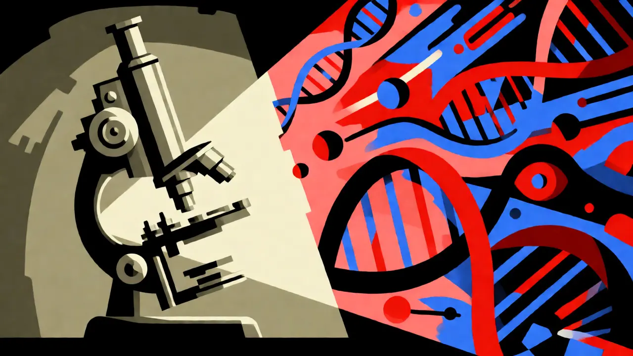
Imagine waking up with a headache that won’t quit, or noticing your vision blurring in one eye. You go to the doctor, get an MRI, and hear words that change everything: "You have a brain tumor." The next question is usually, "What kind? How bad is it?" For decades, doctors answered this by looking at cells under a microscope. Today, the answer is far more complex-and precise. Brain tumors are not just lumps of tissue; they are distinct biological entities defined by both their appearance and their genetic makeup. If you or a loved one are facing this diagnosis, understanding the difference between types, grades, and the new molecular landscape is crucial for navigating treatment options.
The Evolution from Microscope to Molecular Map
For most of the last century, classifying a brain tumor was a visual exercise. Pathologists looked at how abnormal the cells appeared compared to normal brain tissue. This led to the famous grading system we still use today, ranging from Grade 1 (least aggressive) to Grade 4 (most aggressive). But here’s the twist: two tumors that look identical under a microscope can behave completely differently. One might grow slowly for years; another might spread aggressively within months.
This discrepancy drove a major shift in medicine. In 2021, the World Health Organization released the fifth edition of its classification for central nervous system tumors, known as WHO CNS5. This update didn't just tweak the old rules; it rewrote them by integrating molecular diagnostics into the core definition of every tumor type. Now, a diagnosis isn't just "astrocytoma, grade 3." It's "Astrocytoma, IDH-mutant, CNS WHO grade 3." That small addition of "IDH-mutant" changes everything about prognosis and treatment strategy.
Decoding Tumor Grades: What Numbers Really Mean
When you hear "Grade 2," don't think it means a 20% survival rate or a mild case. The grading system describes how the tumor behaves biologically, not necessarily how much time you have left. Think of it as a speedometer rather than a fuel gauge.
- Grade 1: These are slow-growing, often benign tumors. They usually have well-defined edges, making them easier to remove surgically. Pilocytic astrocytomas are a common example, frequently seen in children.
- Grade 2: These are low-grade but infiltrative. The cells look slightly abnormal and tend to spread into surrounding healthy brain tissue. While they grow slowly, they have the potential to recur as higher-grade tumors over time.
- Grade 3: Often called "anaplastic," these tumors show active cell division and invasion into nearby tissue. They are malignant and require more aggressive intervention.
- Grade 4: The most aggressive category. These tumors multiply rapidly, create new blood vessels to feed themselves, and often contain areas of dead tissue (necrosis). Glioblastoma is the most common Grade 4 tumor in adults.
It’s important to note that the WHO CNS5 introduced "within-tumor-type" grading. Previously, all high-grade tumors were lumped together somewhat broadly. Now, grading is specific to the tumor's identity. For instance, oligodendrogliomas only exist as Grade 2 or 3, while meningiomas range from Grade 1 to 3. This precision helps doctors tailor treatments more accurately.

The Molecular Revolution: Why Genetics Matter More Than Looks
If you're undergoing testing, you might wait longer than expected for results-sometimes 7 to 10 business days. This delay happens because labs aren't just counting cells anymore; they are sequencing DNA. Key markers like IDH mutations, genetic alterations in the isocitrate dehydrogenase enzyme that significantly improve prognosis when present, and 1p/19q codeletions determine the true nature of the tumor.
Consider glioblastoma. Historically, any fast-growing, necrotic glioma was called glioblastoma. Under WHO CNS5, if a tumor looks like a glioblastoma but has an IDH mutation, it is reclassified as "Astrocytoma, IDH-mutant, CNS WHO grade 4." This distinction matters because IDH-mutant tumors respond better to treatment and have a median survival of around 31 months, compared to 14.6 months for IDH-wildtype glioblastomas treated with standard care.
| Tumor Type | Required Molecular Markers | Possible Grades | Typical Prognosis Indicator |
|---|---|---|---|
| Glioblastoma | IDH-wildtype | Grade 4 only | Aggressive; median survival ~14.6 months with standard Stupp protocol |
| Astrocytoma | IDH-mutant | Grades 2, 3, 4 | Better response to therapy; longer survival times |
| Oligodendroglioma | IDH-mutant AND 1p/19q-codeleted | Grades 2, 3 | Highly responsive to chemotherapy and radiation |
| Meningioma | Histology-based (mostly) | Grades 1, 2, 3 | Often curable with surgery if Grade 1 |
Multimodal Treatments: Combining Forces Against Cancer
There is no single "cure-all" for brain tumors. Because the brain is protected by the blood-brain barrier-a selective filter that keeps toxins out-it also keeps many drugs out. This necessitates a multimodal approach, combining three main pillars: surgery, radiation, and systemic therapy.
Surgery is usually the first step. The goal is maximal safe resection. Surgeons aim to remove as much of the tumor as possible without damaging critical functions like speech or movement. For some Grade 1 tumors, complete removal can be curative. For higher grades, surgery reduces the tumor burden, making other treatments more effective.
Radiation therapy follows surgery for most malignant tumors. Advanced techniques like stereotactic radiosurgery allow doctors to deliver high doses of radiation precisely to the tumor site while sparing healthy tissue. Systemic therapies include chemotherapy agents like temozolomide, which is the standard for glioblastoma, and newer targeted drugs.
A recent breakthrough illustrates the power of molecular targeting. In June 2023, the FDA accelerated approval of vorasidenib, a drug specifically for IDH-mutant grade 2 gliomas. Clinical trials showed it doubled progression-free survival compared to placebo. This is a game-changer because it delays the need for harsher treatments like radiation in younger patients, potentially preserving cognitive function for longer.
Navigating the Diagnosis: Practical Steps for Patients
Receiving a diagnosis is overwhelming. Many patients report confusion about what the numbers mean. A study found that nearly half of surveyed patients misunderstood grading terminology. To help, here is a practical checklist for your next neuro-oncology appointment:
- Ask for the Integrated Diagnosis: Ensure your report includes both histological features and molecular markers (e.g., IDH status, MGMT promoter methylation).
- Clarify the Grade: Ask specifically if the grade applies to the whole tumor or if there are mixed areas.
- Discuss Treatment Sequencing: For lower-grade tumors, ask if immediate radiation is necessary or if monitoring and targeted therapy are options.
- Inquire About Clinical Trials: Given the rapid evolution of molecular therapies, trials often provide access to cutting-edge treatments before they are widely available.
Costs can also be a concern. Comprehensive molecular testing adds thousands of dollars to diagnostic bills, but insurance coverage is improving as these tests become standard of care. Always verify what your plan covers regarding genomic profiling.
The Future Is Personalized
We are moving away from one-size-fits-all protocols. The integration of liquid biopsies-detecting tumor DNA in cerebrospinal fluid-is showing promise for less invasive monitoring. With sensitivity rates reaching 89% in recent studies, this could reduce the need for repeated surgeries to check for recurrence.
While the statistics for high-grade tumors remain challenging, the trend line is upward. Better classification leads to better trials, which lead to better drugs. Understanding your specific tumor type and grade empowers you to participate actively in your care, asking the right questions and seeking the most appropriate interventions.
Does a Grade 2 brain tumor mean I have cancer?
Not exactly. Grade 2 tumors are considered low-grade gliomas. They are neoplasms (abnormal growths) that are infiltrative and can progress to higher grades, but they are not typically classified as "malignant" in the same aggressive sense as Grade 3 or 4 cancers. However, they do require careful monitoring and often treatment.
Why does my pathology report mention IDH mutation?
The IDH mutation is a key genetic marker that defines certain types of gliomas. Its presence generally indicates a better prognosis and influences treatment choices. Under the current WHO CNS5 guidelines, knowing your IDH status is essential for an accurate diagnosis and determining whether you might benefit from targeted therapies like vorasidenib.
How long does it take to get final brain tumor test results?
Because modern diagnosis requires both microscopic analysis and molecular genetic testing, it typically takes 7 to 10 business days after a biopsy to receive the final integrated diagnosis. This waiting period allows neuropathologists to correlate histological findings with genetic data for the most accurate classification.
Is surgery always required for brain tumors?
Surgery is the primary treatment for most accessible brain tumors to relieve pressure and obtain tissue for diagnosis. However, for some deep-seated or diffuse tumors, or certain low-grade lesions, doctors may recommend close monitoring (watchful waiting) or non-surgical treatments like radiation or medication, depending on the tumor's location and behavior.
What is the difference between glioblastoma and astrocytoma?
Historically, these terms overlapped. Now, under WHO CNS5, "Glioblastoma" refers specifically to IDH-wildtype Grade 4 astrocytic tumors. If an astrocytic tumor has an IDH mutation, it is called "Astrocytoma, IDH-mutant," even if it is Grade 4. This distinction is critical because IDH-mutant tumors have different survival rates and treatment responses.






Amy B September 3, 2026
Whoa, this is actually super helpful. I had no idea the WHO updated their classification in 2021 to include molecular markers like IDH status. It feels like we're finally moving away from just looking at cells under a microscope and really understanding the biology behind the tumor.
The part about vorasidenib delaying radiation for younger patients is huge. Preserving cognitive function is such an underrated aspect of cancer treatment, especially for people who are still working or raising families. Thanks for breaking down the grades so clearly; the speedometer analogy made way more sense than what my doctor explained in ten seconds flat.
Adam Viruet September 3, 2026
Great... another article trying to complicate simple things with fancy words. Grade 1 is slow, Grade 4 is fast. Why do we need "IDH-mutant" and "1p/19q-codeleted" jargon? It's just medical gatekeeping to make doctors feel smarter than patients!!!
Also, "maximal safe resection" sounds nice, but have you ever tried getting brain surgery?? The risks alone are enough to give anyone nightmares. I bet half these "breakthroughs" are just pharma companies trying to sell us new drugs that cost a fortune while doing nothing for the actual quality of life. Just saying...
Jenn Bell September 5, 2026
I think it's important to remember that complexity often equals precision in medicine. While the terminology can be overwhelming, knowing the specific genetic makeup allows for targeted therapies that weren't possible before. For someone facing a diagnosis, having options beyond just "cut it out and burn it" is genuinely hopeful news.
Kimberley Odish September 6, 2026
While your enthusiasm is noted, Adam, your dismissal of molecular diagnostics is intellectually lazy and frankly dangerous for patients relying on accurate prognostication. To suggest that visual grading alone suffices ignores decades of oncological data proving that histology is an imperfect proxy for biological behavior.
Furthermore, labeling standard-of-care genomic profiling as "gatekeeping" demonstrates a profound misunderstanding of how personalized medicine reduces trial-and-error toxicity. If one cannot grasp the distinction between an IDH-wildtype glioblastoma and an IDH-mutant astrocytoma grade 4, one has no business critiquing the necessity of the test. The survival differential is not a marketing gimmick; it is statistically significant clinical reality.
Eric Schultze September 6, 2026
We must consider who benefits from this shift to molecular testing. The insurance companies love this because they can deny coverage for "experimental" treatments until the specific marker is confirmed, yet they charge premiums for tests that may not change immediate surgical intervention.
Moreover, the FDA accelerated approval process is becoming a rubber stamp for pharmaceutical giants. Vorasidenib was approved based on progression-free survival, which is a surrogate endpoint, not overall survival. We are being used as guinea pigs in a system where profit motives drive the definition of "standard care." One must remain skeptical of rapid changes in classification systems when they conveniently align with new product launches.
Jeremy Westcott September 7, 2026
Dude, you're missing the forest for the trees. Before this "molecular revolution," we were basically guessing games with our skulls. Like, literally throwing darts at a board blindfolded. Now we know if the engine is broken (IDH) or if the whole car is on fire (wildtype).
It ain't just pharma greed. My cousin had a glioma that looked nasty under the scope but turned out to be IDH mutant. He got the targeted drug instead of full-blown radiation hell and he's still kicking. That's real. Don't let cynicism cloud the fact that science is actually saving skins here. The conspiracy theorists always want everything to be a scam, but sometimes the tech just works.
Somnath Thombre September 8, 2026
hey guys, interesting read. i had a friend who went through this exact thing last year. the waiting period for results was brutal for him. he kept googling every symptom and driving himself crazy.
the checklist at the end is gold though. asking for the integrated diagnosis saved his family a lot of confusion later on. also, regarding costs, in india we see similar issues with insurance not covering advanced genomic tests fully. its a global problem. hope everyone gets clarity soon
Kim Pender September 9, 2026
Simple point: most people don't read past the first paragraph. They hear "brain tumor" and panic. The grade matters less than the location and whether it's removable. Stop overcomplicating it with genetics if the surgeon can just take it out.
Shu Chowdhury September 10, 2026
That's a fair perspective Kim, but sometimes location makes removal impossible without causing severe deficits. In those cases, the biology tells us if we should watch it closely or hit it hard immediately. It's not about ignoring the surgeon, it's about timing the next steps correctly.
Pramay Dattani September 12, 2026
In India we have world class neurosurgeons who handle complex cases better than many western hospitals. The technology is there. But yes the cost barrier is real. We need govt schemes to cover these molecular tests otherwise only rich people get precise diagnosis. Poor patients suffer due to lack of info